Cartilage Restoration | Advanced Joint Preservation Treatments
Learn about advanced cartilage restoration techniques to relieve joint pain, restore mobility, and prevent arthritis progression. Explore treatment options, benefits, risks, and recovery guidance from expert orthopedic specialists.

Cartilage restoration is a medical procedure designed to repair or regenerate damaged cartilage, which is the flexible connective tissue found in various parts of the body, such as the knee, hip, and shoulder. Cartilage plays a critical role in cushioning joints and enabling smooth movement. When cartilage is damaged due to injury, overuse, or age, it can lead to joint pain, stiffness, and difficulty in movement. Cartilage restoration aims to relieve these symptoms and restore the function of the affected joint.

Early Detection Saves Lives
Early detection and treatment are crucial for improving the chances of survival. If you notice any concerning symptoms, consult a healthcare provider immediately.
Signs and Symptoms

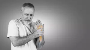
Pain
Persistent or intermittent pain, especially when moving or bearing weight on the affected joint. The pain may worsen after activity or at the end of the day.

Swelling
Fluid buildup around the joint due to inflammation or irritation of the cartilage.

Stiffness
Difficulty bending, straightening, or rotating the joint. The joint may feel tight or "locked."

Limited Range of Motion
Decreased ability to move the joint through its full range due to pain or mechanical obstruction from damaged cartilage.

Popping or Grinding Sounds
A feeling of grinding or clicking in the joint, often referred to as crepitus, which occurs when the cartilage surface is no longer smooth.

Instabilit
The joint may feel weak or "give way" under pressure, especially if the cartilage damage is severe.
Blood in Urine
Hematuria - pink, red, or dark urine, the most common symptom
Frequent Urination
Feeling the need to urinate frequently, even when bladder is not full
Painful Urination
Experiencing pain or burning sensation while urinating
Back or Pelvic Pain
Pain that occurs as the cancer grows and spreads
Unexplained Weight Loss
Significant weight loss not related to diet or exercise
Fatigue
Feeling unusually tired or weak without a clear cause
Meet Our Expert Cartilage Restoration Specialists
Risk Factors
Smoking
Smoking is one of the leading causes of bladder cancer. Chemicals in tobacco smoke can damage the lining of the bladder, increasing the risk.

Gender
Men are at a higher risk of developing bladder cancer than women.
Chronic Bladder Infections or Inflammation
Conditions such as bladder infections and long-term bladder inflammation can increase the risk.

Exposure to Chemicals
Prolonged exposure to certain chemicals, especially those used in the dye industry, rubber production, and chemical manufacturing, increases the risk.

Age
As we age, the cartilage in our joints naturally wears down, becoming thinner and more prone to damage. Older adults are more likely to experience cartilage degradation.
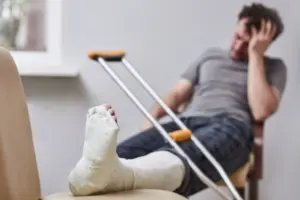
Previous Injury
Past joint injuries, such as fractures or ligament tears, can damage cartilage and lead to further degeneration over time.
Overuse
Repetitive movements or excessive use of a joint (such as in athletes or labor-intensive jobs) can wear down the cartilage.

Obesity
Excess body weight places additional stress on weight-bearing joints, increasing the risk of cartilage damage, particularly in the knees and hips.

Genetics
Some individuals may be genetically predisposed to developing cartilage degeneration, especially conditions like osteoarthritis.

Gender
Women may be at a higher risk of cartilage degeneration due to hormonal changes, especially after menopause.

Poor Posture
Bad posture or improper mechanics during physical activity can lead to uneven pressure on the joints, increasing the likelihood of cartilage damage.

Advanced Cancer Treatment Options
Diet and Nutrition
Prevention
Diagnosis
Key Services
Key Facilities
- Collagen-Rich Foods: Collagen is a key component of cartilage. Bone broth, chicken skin, and fish with skin (like salmon) are good sources of collagen to help promote cartilage health.
- Omega-3 Fatty Acids: These anti-inflammatory fatty acids, found in fish like salmon, mackerel, and sardines, can help reduce joint inflammation and protect cartilage from further damage.
- Vitamin C: Essential for collagen synthesis, Vitamin C supports cartilage repair. Include citrus fruits, bell peppers, broccoli, and strawberries in your diet to meet your Vitamin C needs.
- Vitamin D and Calcium: Both are important for maintaining healthy bones, which support cartilage. Dairy products, fortified plant-based milk, and sunlight are great sources.
- Glucosamine and Chondroitin: These natural compounds, often found in supplements, help support cartilage structure and may aid in the repair of damaged cartilage.
- Turmeric and Ginger: Both have anti-inflammatory properties that can help reduce joint pain and swelling. Add them to your diet for natural pain relief.
- Water: Staying hydrated is essential for joint lubrication and cartilage health, as water helps maintain the elasticity of cartilage and joint fluid.
- Maintain a Healthy Weight: Reducing excess weight decreases the load on weight-bearing joints, especially the knees and hips, reducing the risk of cartilage breakdown.
- Strengthening Exercises: Focus on building strong muscles around the joint to reduce the strain on cartilage. Stronger muscles provide support, which can prevent cartilage wear.
- Use Proper Technique: When exercising or engaging in physical activities, using proper form and technique can help prevent unnecessary stress on joints.
- Low-Impact Exercises: Engage in low-impact exercises such as swimming, cycling, and walking to protect joints from excessive wear and tear.
- Stretch and Warm-Up: Stretching and warming up before physical activity help prepare joints and muscles for movement, reducing the risk of injury and cartilage damage.
- Avoid Overuse: Take regular breaks from repetitive activities to reduce the risk of joint strain. Rest allows cartilage to heal and recover from minor injuries.
- Use Joint Protection: Wear protective gear, such as knee pads or wrist guards, during high-impact sports to protect cartilage from sudden trauma.
- Physical Examination: The doctor will assess the joint’s range of motion, tenderness, swelling, and signs of instability. They may also test for mechanical issues such as clicking or grinding.
- X-rays: X-rays help rule out bone fractures or joint deformities but do not provide detailed images of cartilage. However, they can show signs of joint degeneration and bone spurs.
- MRI (Magnetic Resonance Imaging): An MRI provides high-resolution images of soft tissues, including cartilage, making it the gold standard for diagnosing cartilage damage.
- Arthroscopy: In some cases, a minimally invasive procedure using a small camera inserted into the joint (arthroscope) is used to directly visualize the cartilage and assess the extent of damage.
- Ultrasound: Sometimes used to examine the soft tissue around the joint and detect fluid buildup or other abnormalities.
- Conservative Treatments: Non-surgical options such as physical therapy, anti-inflammatory medications, corticosteroid injections, or hyaluronic acid injections can help reduce pain and improve joint mobility.
- Stem Cell Therapy: A newer treatment option that involves injecting stem cells into the joint to promote the regeneration of damaged cartilage.
- Platelet-Rich Plasma (PRP) Therapy: Involves injecting concentrated platelets from your blood into the affected joint to promote healing and cartilage regeneration.
- Microfracture Surgery: A minimally invasive surgical technique where tiny holes are made in the bone beneath the damaged cartilage to stimulate the growth of new cartilage.
- Autologous Chondrocyte Implantation (ACI): This procedure involves taking cartilage cells from the patient’s own body, growing them in a lab, and re-implanting them into the damaged area.
- Osteochondral Grafting: Involves transferring healthy cartilage from another part of the body or using cadaver cartilage to replace damaged cartilage.
- Joint Replacement Surgery: In severe cases of cartilage damage, joint replacement surgery may be considered to replace the damaged joint with a prosthetic one.
- Orthopedic Centers: Specialized centers that focus on joint health and cartilage restoration, offering a variety of treatments, including physical therapy, injections, and surgery.
- Sports Medicine Clinics: Clinics that focus on preventing, diagnosing, and treating injuries related to physical activity, offering advanced treatments like stem cell therapy and PRP.
- Rehabilitation Centers: Post-treatment rehab facilities equipped with physical therapists experienced in joint recovery and cartilage restoration, focusing on improving mobility and reducing pain.
- Surgical Suites: Hospitals or surgical centers with state-of-the-art equipment for performing minimally invasive and advanced cartilage restoration surgeries, including arthroscopic procedures.
- Diagnostic Imaging Centers: Facilities that offer advanced imaging techniques such as MRI, X-rays, and ultrasounds to accurately diagnose cartilage damage.
Top Medical Facilities at Our Multispeciality Hospital – Here’s What Makes Us Different!
Ready to Begin Your Cartilage Restoration Journey?
Learn More About Cartilage Restoration Care
Frequently Asked Questions
Success rates vary based on the severity of the cartilage damage, age, and overall health of the patient. Generally, microfracture and ACI surgeries show good results, with many patients experiencing significant improvement in pain and joint function.
Recovery time depends on the procedure used. For minimally invasive treatments like microfracture, recovery can take 4-6 months, while more complex surgeries like ACI may take 9-12 months for full recovery.
While cartilage restoration treatments can be effective, there are risks such as the possibility of incomplete healing, re-injury, or joint instability. Long-term risks may vary based on the treatment approach, but proper rehabilitation reduces the likelihood of complications.