Advanced Ultrasound Services | Accurate Diagnostic Imaging
Get advanced ultrasound services with high-resolution imaging and expert radiologists for precise diagnosis, early detection, and effective treatment planning.
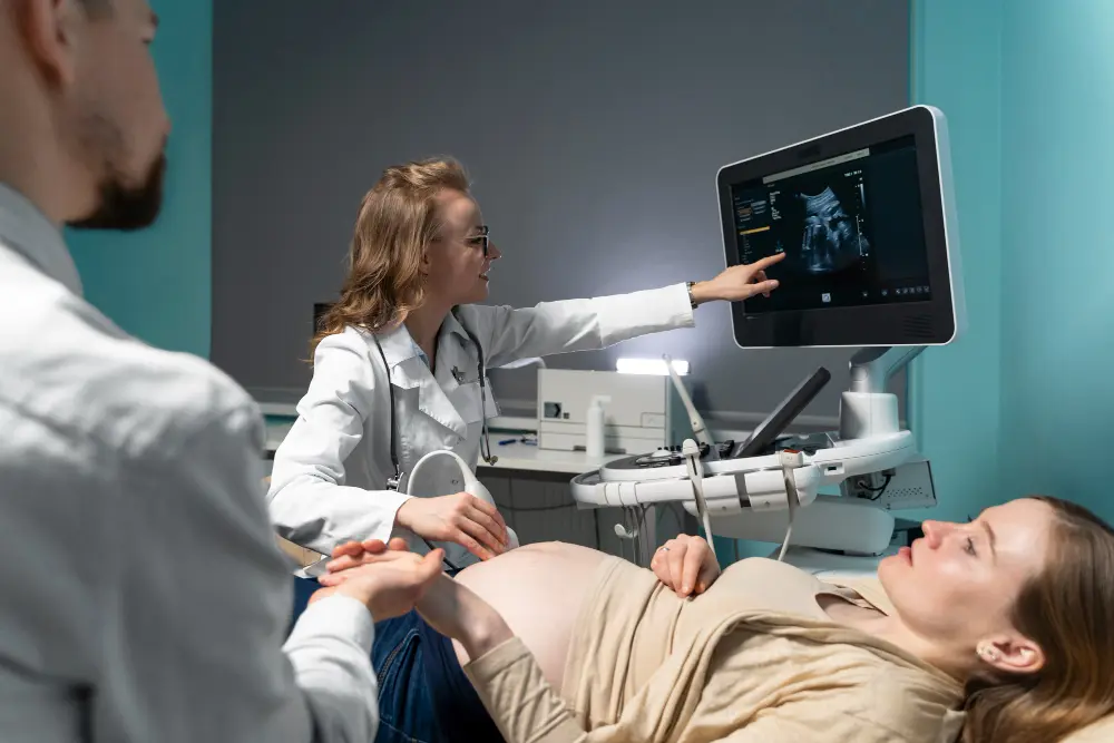
Ultrasound, also known as sonography, is a non-invasive imaging technique that uses high-frequency sound waves to create real-time images of the body’s internal organs and tissues. It’s one of the safest diagnostic tools in modern medicine no radiation, no pain, and no downtime. From tracking a baby’s growth during pregnancy to identifying gallstones or heart issues, ultrasound plays a vital role in accurate and early diagnosis. It provides instant results, helping doctors plan the right treatment without unnecessary delays.
2-15 Mhz
Ultrasound for diagnostic usage produces frequency range of 2-15 Mhz
2.5 Mhz
2.5 Mhz frequencies are used for deep abdomen, gynecological and obstetric imaging
3.5 Mhz
3.5 Mhz frequencies for general abdomen, gynecological and obstetric imaging
How Ultrasound Works
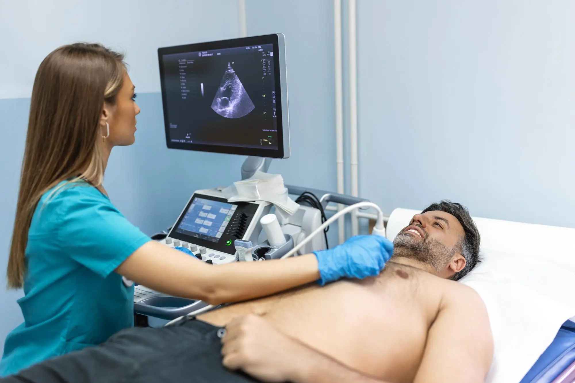
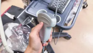
The process is powered by sound waves that bounce off the body’s internal structures. A small handheld device called a transducer emits these sound waves, which then reflect back to create detailed images on a screen.
A conductive gel is applied to the skin to eliminate air pockets and ensure smooth transmission of waves. The reflected sound patterns are processed by a computer to form visual representations of organs, tissues, and blood flow in real time. Different types of ultrasounds abdominal, pelvic, vascular, echocardiogram, and obstetric are chosen based on the part of the body being examined.
Why Ultrasound is Done
Ultrasound isn’t just about pregnancy scans it’s a versatile diagnostic tool. Doctors use it for various reasons, including
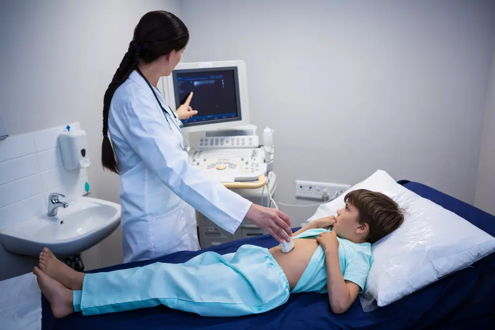
Examining abdominal pain
Detecting gallstones, liver diseases, or kidney blockages.
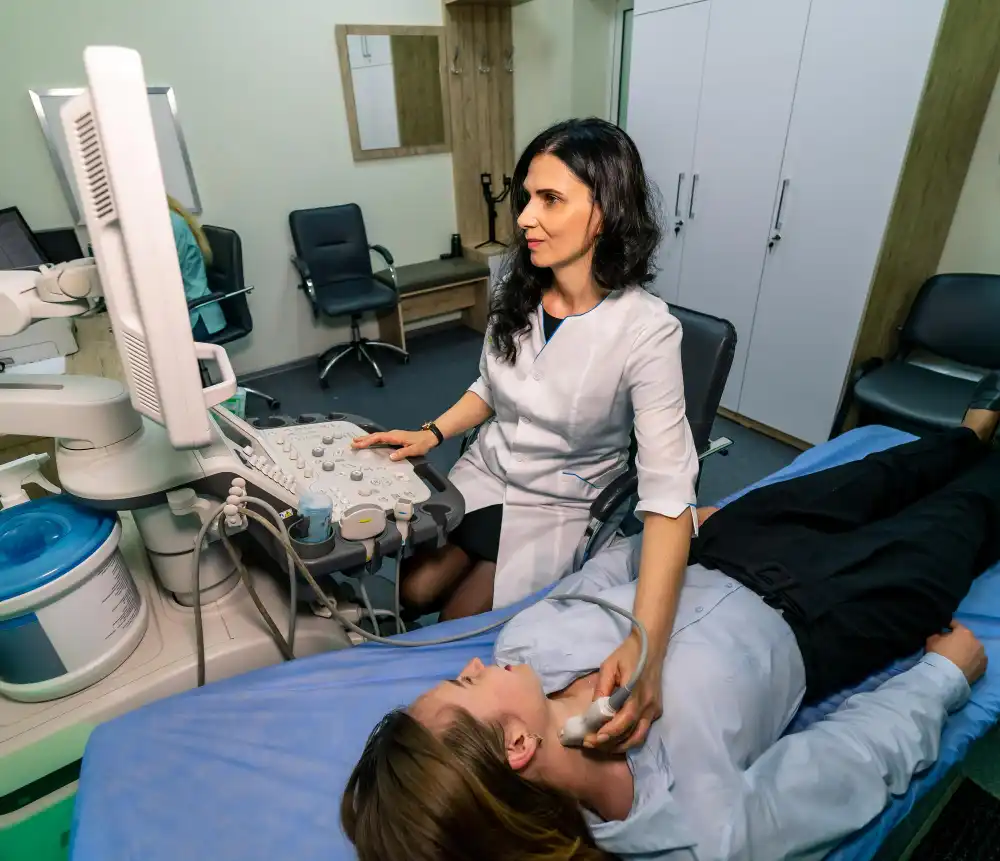
Checking blood flow
Identifying clots, narrowing arteries, or vascular disorders.
Evaluating soft tissues
Spotting cysts, lumps, or tumors in organs or glands.
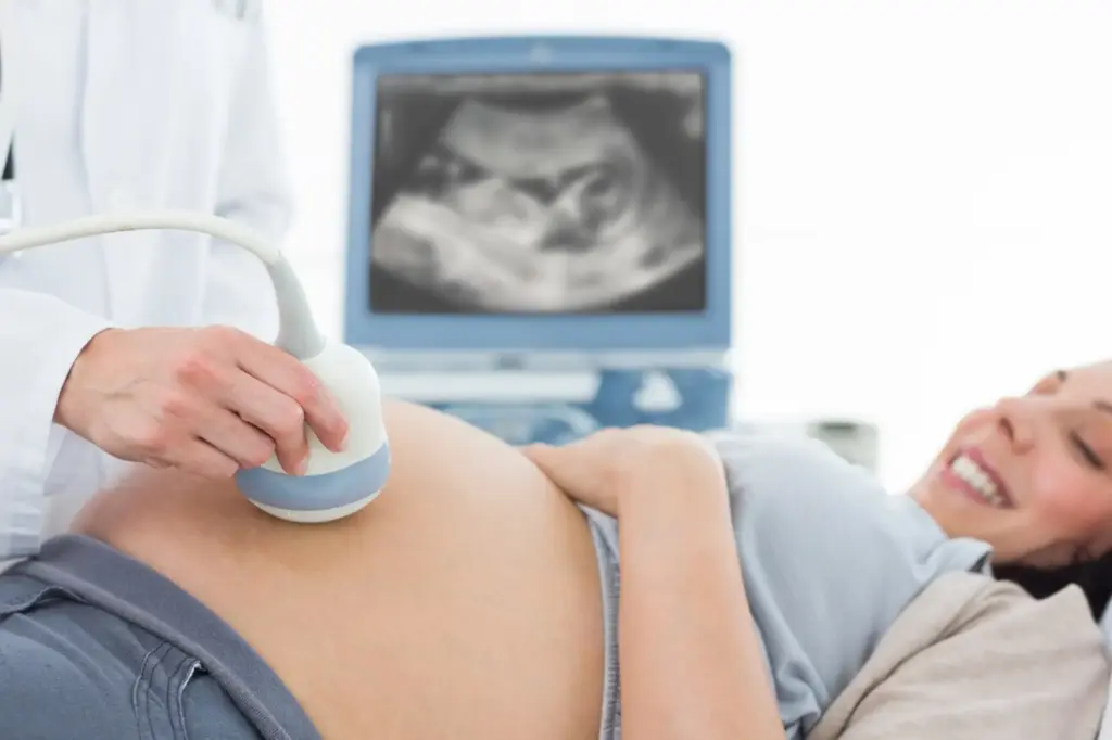
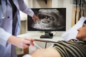
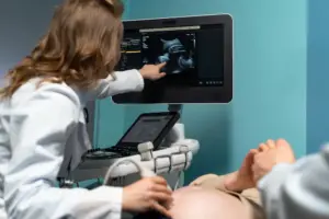
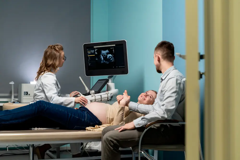
Monitoring pregnancy
Observing fetal growth, position, and heartbeat.
Guiding medical procedures
Such as biopsies or fluid drainage with precision.
Benefits of Ultrasound
Radiation-Free
Unlike X-rays or CT scans, ultrasound uses harmless sound waves perfect for children and pregnant women.
Real-Time Imaging
It provides instant visualization of organs and blood flow, making it ideal for emergency diagnostics.
Non-Invasive & Painless
No injections, no discomfort just a quick scan with a cool gel.
Portable and Affordable
Ultrasound machines are compact and cost-efficient, allowing scans even at bedside or in clinics.
Helps Early Detection
Subtle abnormalities can be caught early, improving treatment outcomes dramatically.
Preparation Before an Ultrasound
Preparation depends on the type of ultrasound being performed:
Abdominal Ultrasound: Patients are usually advised to fast for 6–8 hours before the scan to reduce gas and improve clarity.
Pelvic or Obstetric Ultrasound: A full bladder is essential to get clear images, so patients are asked to drink plenty of water before the test.
Vascular or Soft Tissue Ultrasound: Usually requires no special preparation.
It’s always best to follow the exact instructions given by the radiology team to ensure the most accurate results.
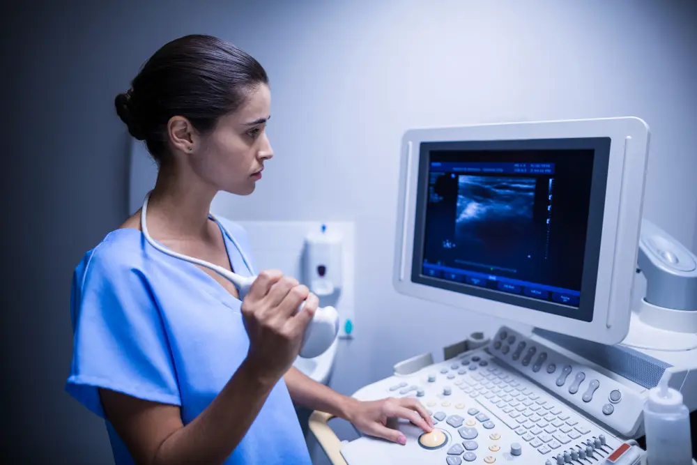
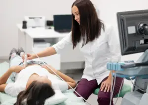
During the Procedure
An ultrasound is simple and comfortable:
You’ll be asked to lie on an examination table.
A clear, water-based gel is applied to the area being scanned.
The technician moves the transducer gently over your skin.
Images instantly appear on the monitor, allowing real-time observation.
Depending on the type of ultrasound, the procedure takes anywhere from 10 to 30 minutes. For pregnancy scans, you may even get to see your baby’s first movements or heartbeat a moment most parents cherish forever.
After the Procedure
Once the scan is done, the gel is wiped off and you can resume normal activities immediately there’s no recovery time needed. A radiologist will analyze the captured images and send a report to your doctor. Based on this, your doctor may discuss next steps, such as further imaging or treatment. In emergency cases, results are often available right away.